Robotic Bronchoscopy
Overview
Robotic bronchoscopy, also known as robotic-assisted bronchoscopy, is a recent advancement in bronchoscopy, the procedure used to biopsy lung nodules to detect the presence of lung cancer and other lung diseases.
Lung cancer is the third most common type of cancer and the leading cause of cancer-related death in the United States. Early diagnosis and treatment often lead to better outcomes.
A diagnosis of lung cancer often begins when a chest X-ray or computed tomography (CT) scan shows a nodule—an area of abnormal tissue—in the lungs. If the nodule is suspicious or grows over time, doctors will perform a biopsy to collect a tissue sample that can be tested for the presence of cancer cells.
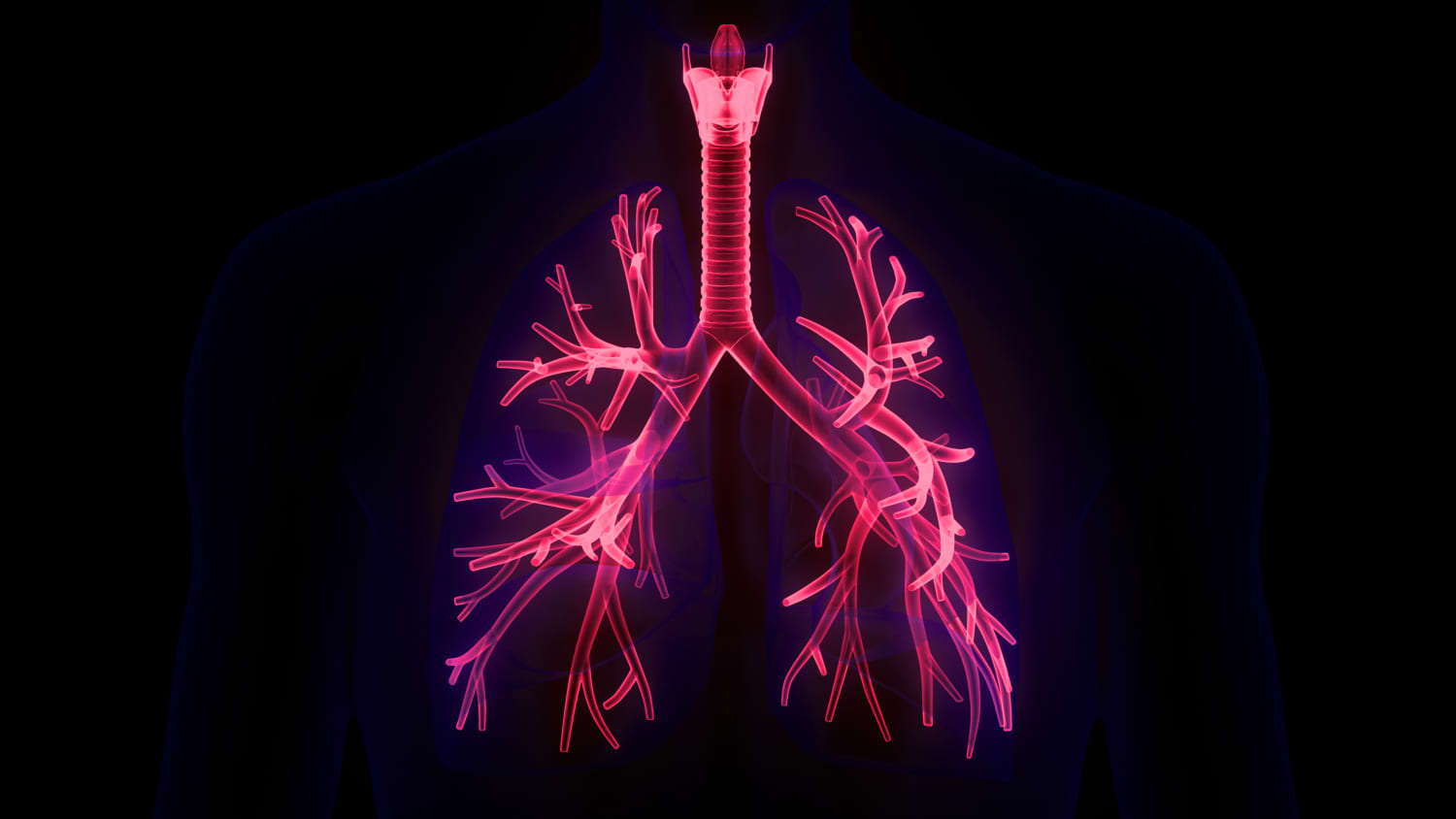
Bronchoscopy is a widely used procedure for biopsying lung nodules. In traditional bronchoscopy, a doctor manually guides a thin tube, called a bronchoscope, into the patient’s mouth or nose, down the throat, past the vocal cords and windpipe, and into the passageways of the lungs. The bronchoscope is equipped with a light, camera, and biopsy tools that allow doctors to visually examine and biopsy nodules.
But robotic bronchoscopy is different. Like traditional bronchoscopy, it’s a minimally invasive procedure that allows doctors to biopsy nodules in the lungs. The difference is that in robotic bronchoscopy, the doctor uses a controller at a console to operate a robotic arm. The robotic arm guides a catheter—a thin, flexible, and maneuverable tube equipped with a camera, light, and shape-sensing technology—through the patient’s airways.
The robotic arm’s precise movements enable doctors to accurately direct the catheter around tight turns in the airways and into the hard-to-reach areas of the lungs. This means doctors can examine and biopsy suspicious nodules—and potentially detect cancer—in parts of the lungs that may be inaccessible with traditional bronchoscopy. What’s more, the procedure is safe—serious complications are rare—and recovery is usually quick.
"As part of the comprehensive Center for Thoracic Cancers, we are now able to offer patients the option of robotic bronchoscopy,” says Yale Medicine interventional pulmonologist Christopher Morton, MD. “This technology will allow us to biopsy lung nodules and masses with improved accuracy and fewer side effects, in addition to lymph node biopsies that we already do. This will get patients diagnosed and referred to the appropriate treating physician quicker."
Who is eligible for robotic bronchoscopy?
People who have a lung nodule or mass that shows signs of growth or is otherwise suspicious may be eligible for robotic bronchoscopy. To determine if an individual can receive the procedure, doctors will review their medical history and perform a physical exam to assess their overall health and lung and heart function. Blood tests to evaluate kidney function and measure blood-clotting factors, among others, may also be used. The purpose of the exam and tests is to rule out medical conditions that may increase the risk of complications during the procedure.
Those who will benefit from robotic bronchoscopy will have CT scans of their chest done before the procedure. These scans allow doctors to locate nodules within the airway. The robotic bronchoscopy system then produces a virtual three-dimensional (3D) image of the lungs and the airways within the lungs from the CT scans. The system’s planning software creates virtual pathways through the airways to nodules in the lungs. During the procedure, doctors guide the robotic bronchoscope through airways alongside with these virtual pathways to examine and biopsy nodules.
What happens during robotic bronchoscopy?
During robotic bronchoscopy, you will be under general anesthesia, which means you will be unconscious and not feel any pain during the procedure. An endotracheal tube will be placed in your windpipe to keep the airway open and provide oxygen.
The doctor then uses a traditional flexible bronchoscope to examine your airway. Next, the doctor docks the robotic bronchoscope system into the endotracheal tube. Using a controller at a console, a doctor maneuvers the robotic arm and catheter. Using the 3D image of the airways (generated from CT scans), the doctor directs the catheter down the throat and through the airways in the lung.
The catheter comes with “shape-sensing” technology that can communicate its location in the lungs to an external monitor. When the catheter reaches the target lung nodule, biopsy tools, such as a fine needle or forceps, will be passed through it and used to collect tissue samples from the nodule. Usually, several tissue samples are collected from different parts of each nodule. Doctors may also use 3D fluoroscopy for real-time imaging during the procedure to improve accuracy of lung nodule localization and biopsy.
In many cases, following biopsy of the nodule, doctors may perform another procedure known as endobronchial ultrasound, or EBUS, to biopsy lymph nodes around the lungs and airways. This helps not just with the diagnosis but also with the staging of lung cancer. Performing robotic-assisted bronchoscopy and EBUS under single anesthesia will help reduce the unnecessary waiting times and anxiety that patients may traditionally experience.
What happens after the procedure?
After the procedure, you will be closely monitored as the effects of anesthesia wear off. You may have a sore throat and shouldn’t eat or drink anything for one to four hours after the procedure. An hour or longer after the procedure, you may have a chest X-ray to rule out a collapsed lung, which is a possible complication of the procedure.
Robotic bronchoscopy is typically an outpatient procedure, meaning you can usually go home the same day as the procedure.
What are the risks of robotic bronchoscopy?
Robotic bronchoscopy is a safe procedure, but there is a small risk for certain complications. Serious complications are rare.
Complications may include:
- Bleeding
- Collapsed lung (known as pneumothorax)
- Airway perforation
- Temporary bronchospasm (tightening of the muscles of the airways in the lungs, making it harder to breathe)
- Temporary laryngospasm (tightening of the muscles of the vocal cords, making it harder to speak and/or breathe)
- Bloody mucus (for the first day or two after the procedure)
- Hypoxemia (lower than normal level of oxygen in the blood)
- Sore throat
- Hoarseness
- Nausea
- Fever
- Cough
- Heart arrhythmia
- Infection
What stands out about Yale Medicine's approach to robotic bronchoscopy
“Our highly trained interventional pulmonologists and thoracic surgeons will be performing robotic bronchoscopy at Yale,” says Yale Medicine interventional pulmonologist Sanket Thakore, MD. “They make lung cancer diagnosis and management the main focus of their practice. Additionally, they are on the cutting edge and use the most up-to-date techniques and technology to provide the highest level of care to their patients. For lung cancer patients, they work closely with other experts in the Robotic Bronshoscopy Program to provide the best treatments and outcomes.”